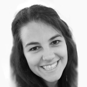
Raquel Conceição

Gabinete no IBEB
1.03
Contactos
E-mail: rcconceicao[at]fc.ul.pt
Redes profissionais
Currículo
Tópicos de investigação
- Aprendizagem automática para aplicações médicas
- Cancro da mama
- Classificação de Tumores
- Desenvolvimento de fantomas anatómicos
- Gânglios linfáticos axilares
- Imagem Médica Por Microondas
- Processamento de Imagem
- Processamento de Sinal
Biografia
Sou professora auxiliar com agregação e investigadora com um histórico significativo de publicações revistas por pares e projetos europeus financiados. Sou a primeiro investigadora doutorada, em Portugal, com doutoramento na área de imagiologia médica por micro-ondas, tendo iniciado os primeiros esforços de investigação nesta área a nível nacional. Fui também a mais jovem coordenadora de uma Ação COST, com a Ação TD1301. Outros financiamentos europeus que obtive incluem uma Marie Curie Intra-European Fellowship (FP7), e uma Marie Skłodowska-Curie Action para uma Innovative Training Networks (H2020), como beneficiária.
Sou vice-presidente do Comité Português da URSI para a Comissão K “Eletromagnetismo em Biologia e Medicina” e Membro do Comité MTT TC-28 Efeitos Biológicos e Aplicações Médicas. Atualmente, sou a coordenadora científica e investigadora no Instituto de Biofísica e Engenharia Biomédica – IBEB, desenvolvendo técnicas de imagiologia por micro-ondas para detetar e classificar o cancro da mama, bem como gânglios linfáticos axilares metastizados, desempenhando um papel importante numa das áreas prioritárias de pesquisa do instituto, “Imagiologia Médica e Diagnóstico”. Tenho outros interesses de investigação que incluem técnicas de modelação com aprendizagem automática, engenharia biomédica, processamento de sinais e imagens, e investigação aplicada de engenharia eletrónica. Sou autora de 34 artigos em revistas científicas, 56 artigos em conferências e editora/autora de 4 livros, e também organizei com sucesso uma “special issue” numa revista de categoria Q1, Sensors. Colaborei e co-autorei com mais de 100 investigadores internacionais.
Desde que me tornei professora auxiliar no Departamento de Física da Faculdade de Ciências da Universidade de Lisboa, Portugal, lecionei 11 disciplinas diferentes. Assumi o papel de regente várias vezes, e dei aulas a alunos de licenciatura e mestrado em Engenharia Biomédica e Biofísica, e outros estudantes do Departamento de Física. Orientei 7 estudantes de doutoramento e 28 estudantes de mestrado, além de outros 28 projetos científicos de licenciatura.
Fui vice-presidente do Departamento de Física na mesma instituição, contribuindo de forma significativa para atividades de divulgação interna e externa, entre 2018 e 2021. Entre 2019 e 2022, fui vice-coordenadora do Mestrado Integrado em Engenharia Biomédica e Biofísica, e em 2021-2022, fui coordenadora do Mestrado em Engenharia Biomédica e Biofísica. Possuo um doutoramento em Engenharia Elétrica e Eletrónica (2011) e um Mestrado Integrado em Engenharia Biomédica (2007), da National University of Ireland Galway (NUIG) e da Faculdade de Ciências e Tecnologia, Universidade Nova de Lisboa – FCTUNL, respetivamente.
GERADOR DE FANTOMAS ANTROPORMÓFICOS DA CABEÇA E PESCOÇO, doi:10.3390/s20072029
Alunos de doutoramento
2022 – a decorrer, Teresa Pereira (PhD in Computer Science), Universidade de Aveiro, “Electrocardiogram for Biometric Recognition: Collectability, Stability and Application Challenges”.
2021 – a decorrer, Ana Sofia Verde (PhD in Biomedical Engineering and Biophysics), Universidade de Lisboa, “Validating biologically meaningful radiomics signatures for prostate cancer”.
2021 – a decorrer, Maria Gonçalves (PhD in Biomedical Engineering and Biophysics), Universidade de Lisboa, “Multi-Scale Modelling of Breast Preservation following Cancer Treatment”.
2020 – a decorrer, Ana Catarina Pelicano (PhD in Biomedical Engineering and Biophysics), Universidade de Lisboa, “Bringing Medical Microwave Imaging of the Breast Closer to Clinical Realism”.
2018 – 2023, Matteo Savazzi (PhD in Biomedical Engineering and Biophysics), Universidade de Lisboa, “Electromagnetic device for axillary lymph node diagnosis”.
2018 – 2022, Daniela Godinho (PhD in Biomedical Engineering and Biophysics), Universidade de Lisboa, “Microwave Imaging to Improve Breast Cancer Diagnosis”.
2012 – 2019, Filipa Borlinhas (PhD in Biomedical Engineering and Biophysics), Universidade de Lisboa, “Diffusion-Weighted Imaging (DWI) in Breast Magnetic Resonance Imaging”
Alunos de mestrado
2023 – a decorrer, Cláudio Jorge, “UterineExplorer Plus – Toolbox for uterine eletromyography processing and visualisation”.
2023 – a decorrer, Raquel Rebordão, “The role of mechanical forces in inhibiting cell proliferation in the heart”.
2023 – a decorrer, Tiago Silva, “Image Reconstruction aided by Machine Learning for Breast Microwave Imaging”.
2022 – a decorrer, Inês André, “Improvement of Machine Learning-based Image Reconstruction Algorithms for Breast Microwave Imaging”.
2021 – a decorrer, Maria Bernardo, “Neuroimaging of Addictive Behaviour”.
2021 – a decorrer, José António da Silva Raimundo, “Design of a hybrid lipid nanosystem combining cytotoxic and magnetic properties”
2021 – 2024, Miguel Fidalgo, “Convolutional Neural Networks in Prostate Cancer Detection, Segmentation and Classification using mpMRI images and feature-selected Radiomic Features”.
2021 – 2023, Pedro Lemos Cidadão, “Medical Microwave Imaging of MRI-based Axillary region models via 3D Finite Difference Time Domain”.
2021 – 2023, Maria Lopes, “Comparing single-shell and multi-shell free-water fraction estimation algorithms”.
2021 – 2023, Catarina Moura, “Reconstruction of Microwave Imaging using Machine Learning”.
2021 – 2023, Luísa Freitas, “PID control of depth of hypnosis in anesthesia for propofol and remifentanil coadministration”
2020 – 2022, Mariana Piedade, “Robustness of photon dose distributions against intra and inter-fraction anatomical changes for whole lung irradiation”.
2021 – 2022, Mariya Berezhanska, “Estudo exploratório do uso de Imagem por Micro-ondas para deteção de cáries dentárias”.
2020 – 2022, Helena Sousa, “A Patch-Wise Generative Adversarial Network for PET-MR Image Generation with Feature Attribution for Detection of Focal Cortical Dysplasias”.
2020 – 2022, Ângelo Nunes, “Optimizing T2 quantitative estimation of knee cartilage with Magnetic Resonance Imaging applying dictionary-based methods”.
2020 – 2022, Cláudia Baleia, “Comparison of T1-maps and Late Gadolinium Enhancement in the detection of myocardial fibrosis”.
2020 – 2021, Teresa Pereira, “Biometric Authentication and Identification through Electrocardiogram Signals”.
2020 – 2021, Carolina Piçarra, “Automatic Reporting of TBI Lesion Location in CT based on Deep Learning and Atlas Mapping”.
2019 – 2021, Madalena Valente, “Deep Learning for Multi-animal Tracking”.
2019 – 2021, Miguel Rodrigues, “Classifying Breast Tumors using Medical Microwave Radar Imaging”.
2019 – 2020, Márcia Abreu, “Computational modeling of TGF-𝛃 signaling”.
2019 – 2020, Luís Venâncio, “Prostate Lesion Segmentation with Convolutional Neural Networks”.
2018 – 2020, Ana Catarina Pelicano, “Modelling the head and neck region for microwave imaging of cervical lymph nodes”.
2018 – 2019, Carolina Seabra, “Prostate cancer biochemical recurrence prediction after radical prostatectomy using machine learning analysis of histopathology”.
2018 – 2019, Nuno Silva, “Translation of medical devices: thermal properties of biological tissues and a nasogastric tube detector device”.
2014 – 2016 (unfinished), Rui Santos, “Detection of Axilla Lymph Nodes with Microwave Imaging to Improve Breast Cancer Diagnosis”.
2014 – 2015, Nádia Vilhena, “Ultrasound assisted oncolytic virotherapy: in vitro and in vivo studies”.
2013 – 2014, Ricardo Eleutério, “Microwave Imaging of the Axilla to Aid Breast Cancer Diagnosis”.
2013 – 2014, Alexandre Medina, “Development of Phantoms for the Axilla and Breast Tumours to Simulate Microwave Imaging”.
2012 – 2013, Luís Oliveira, “Desenvolvimento de modelos numéricos da zona axilar a utilizar em imagem por micro-ondas como complemento ao diagnóstico do cancro da mama” (Development of numerical models of the axillary region for microwave imaging as a diagnostic aid for breast cancer).
2012 – 2013, Hugo Medeiros, “Classificação de Tumores de Cancro na Mama através de Radar de Banda Ultra-Larga de Microondas” (Classification of Breast Cancer Tumours through Microwave Ultra Wideband Radar Imaging).
Projectos (selecção)
2024-2027 (seleccionado para financiamento a 1/2/2024) Project Coordinator of Horizon Europe HORIZON-WIDERA 2023-ACCESS 02-01 Twinning “Bone, Brain, Breast and Axillary Medical Microwave Imaging Twinning (3BAtwin)”, 1.5M€
2023-2024 (a decorrer) Principal Investigator of Exploratory Project by FCT, “First Patient Study for Axillary and Breast Medical Microwave Imaging”, https://doi.org/10.54499/2022.08973.PTDC, 50k€
2019-2024 (a decorrer) Research Unit Coordinator (since July/2022) of FCT Strategic Programme for Portuguese Research Units 2018, https://doi.org/10.54499/UIDB/00645/2020 & https://doi.org/10.54499/UIDP/00645/2020, >500k€
2018-2023 Working Group Leader of COST Action CA17115 MyWAVE, www.cost.eu/actions/CA17115, 523k€
2018-2022 Beneficiary of H2020 MSCA-ITN EMERALD, www.msca-emerald.eu/, 3.3M€
2014-2018 Member of COST Action BM1309, www.cost.eu/actions/bm1309, 654k€
2014-2016 Post-doctoral researcher of EPSRC, “Hybrid UWB Radar/Inverse Scattering Approaches for Breast Cancer Imaging”, http://gow.epsrc.ac.uk/NGBOViewGrant.aspx?GrantRef=EP/J007293/1, 272k£
2013-2017 Chair of COST Action TD1301, www.cost.eu/actions/td1301, 520k€
2013-2015 Fellow of FP7 Marie Curie Intra European Fellowship, BREASTCANCERDETECT, https://cordis.europa.eu/project/rcn/103278_en.html, 151k€
2012 Post-doctoral fellow of FCT “Uma nova metodologia multimodal de imagem para o cancro da mama: PEM-UWB”, 55k€
Publicações
Publicações em revistas
(2024) The Complex Permittivity of Biological Tissues: A Practical Measurement Guideline, IEEE Access 12, p. 10296-10314, url, doi:10.1109/ACCESS.2024.3352728
(2024) In Vivo Measurements of Murine Melanoma's Complex Permittivity between 500 MHz - 8.5 GHz, IEEE Antennas and Wireless Propagation Letters, doi:10.1109/LAWP.2024.3460183
(2024) Repository of MRI-derived models of the breast with single and multiple benign and malignant tumors for microwave imaging research, PLoS ONE 19(5 May), p. e0302974, Public Library of Science San Francisco, CA USA, doi:10.1371/journal.pone.0302974
(2023) Experimental Assessment of Axillary Lymph Node Microwave Tomography Using Anthropomorphic Phantoms, IEEE Journal of Electromagnetics, RF and Microwaves in Medicine and Biology 7(2), p. 160-167, IEEE, doi:10.1109/JERM.2023.3241777
(2023) Dielectric Characterization of Healthy Human Teeth from 0.5 to 18 GHz with an Open-Ended Coaxial Probe, Sensors 23(3), p. 1617, Multidisciplinary Digital Publishing Institute, doi:10.3390/s23031617
(2023) Biometric Recognition: A Systematic Review on Electrocardiogram Data Acquisition Methods, Sensors 23(3), p. 1507, Multidisciplinary Digital Publishing Institute, doi:10.3390/s23031507
(2023) Evaluating the Performance of Algorithms in Axillary Microwave Imaging towards Improved Breast Cancer Staging, Sensors 23(3), p. 1496, MDPI, doi:10.3390/s23031496
(2023) Dielectric Characterization of Healthy Human Teeth from 0.5 to 18 GHz with an Open-Ended Coaxial Probe, Sensors 23(3), p. 1617, MDPI, doi:10.3390/s23031617
(2023) Experimental Assessment of Axillary Lymph Node Microwave Tomography Using Anthropomorphic Phantoms, IEEE Journal of Electromagnetics, RF and Microwaves in Medicine and Biology, IEEE
(2022) Initial Study Using Electrocardiogram for Authentication and Identification, Sensors 22(6), p. 2202, MDPI, doi:10.3390/s22062202
(2022) Experimental Evaluation of an Axillary Microwave Imaging System to Aid Breast Cancer Staging, IEEE Journal of Electromagnetics, RF and Microwaves in Medicine and Biology 6(1), p. 68-76, IEEE, doi:10.1109/JERM.2021.3097877
(2022) Modelling level I Axillary Lymph Nodes depth for Microwave Imaging, Physica Medica 104, p. 160-166, Elsevier, doi:10.1016/j.ejmp.2022.11.008
(2021) Application of machine learning to predict dielectric properties of in vivo biological tissue, Sensors 21(20), p. 6935, Multidisciplinary Digital Publishing Institute, doi:10.3390/s21206935
(2021) Study of Freezing and Defrosting Effects on Complex Permittivity of Biological Tissues, IEEE Antennas and Wireless Propagation Letters 20(12), p. 2210-2214, https://doi.org/10.1109/LAWP.2021.3102842, doi:10.1109/LAWP.2021.3102842
(2021) Evaluation of Refraction Effects in Dry Medical Microwave Imaging Setups, IEEE Antennas and Wireless Propagation Letters 20(4), p. 617-621, IEEE, doi:10.1109/LAWP.2021.3059162
(2021) Development of MRI-based axillary numerical models and estimation of axillary lymph node dielectric properties for microwave imaging, Medical Physics 48(10), p. 5974-5990, Wiley, doi:10.1002/mp.15143
(2021) Evaluation of Refraction Effects in Dry Medical Microwave Imaging Setups, IEEE Antennas and Wireless Propagation Letters 20(4), p. 617-621, IEEE, doi:10.1109/LAWP.2021.3059162
(2021) Development of 3d mri-based anatomically realistic models of breast tissues and tumours for microwave imaging diagnosis, Sensors 21(24), p. 8265, MDPI, doi:10.3390/s21248265
(2021) Development of MRI-based axillary numerical models and estimation of axillary lymph node dielectric properties for microwave imaging, Medical Physics 48(10), p. 5974-5990, Wiley, doi:10.1002/mp.15143
(2020) Development of a 3d anthropomorphic phantom generator for microwave imaging applications of the head and neck region, Sensors (Switzerland) 20(7), p. 2029, MDPI, doi:10.3390/s20072029
(2020) Characterisation of ex vivo liver thermal properties for electromagnetic-based hyperthermic therapies, Sensors (Switzerland) 20(10), p. 3004, MDPI, doi:10.3390/s20103004
(2020) Classification of breast tumor models with a prototype microwave imaging system, Medical Physics 47(4), p. 1860-1870, Wiley Online Library, doi:10.1002/mp.14064
(2019) Gamma Distribution Model in the Evaluation of Breast Cancer Through Diffusion-Weighted MRI: A Preliminary Study, Journal of Magnetic Resonance Imaging 50(1), p. 230-238, doi:10.1002/jmri.26599
(2019) Optimal b-values for diffusion kurtosis imaging in invasive ductal carcinoma versus ductal carcinoma in situ breast lesions, Australasian Physical and Engineering Sciences in Medicine 42(3), p. 871-885, Springer, doi:10.1007/s13246-019-00773-2
(2018) Diagnosing breast cancer with microwave technology: Remaining challenges and potential solutions with machine learning, Diagnostics 8(2), p. 36, Multidisciplinary Digital Publishing Institute, doi:10.3390/diagnostics8020036
(2018) Diagnosing breast cancer with microwave technology: Remaining challenges and potential solutions with machine learning, Diagnostics 8(2), p. 36, Multidisciplinary Digital Publishing Institute, doi:10.3390/diagnostics8020036
(2017) Digital analysis of tumour microarchitecture as an independent prognostic tool in breast cancer, Modern Pathology 30(Supplement 2s), p. 68A, Nature Publishing Group
(2016) Development of Clinically Informed 3-D Tumor Models for Microwave Imaging Applications, IEEE Antennas and Wireless Propagation Letters 15, p. 520-523, IEEE, doi:10.1109/LAWP.2015.2456051
(2015) Other applications of medical microwaves – Breast tumour classification, European Journal of Molecular & Clinical Medicine 2(2), p. 62, No longer published by Elsevier, doi:10.1016/j.nhtm.2014.11.028
(2014) Compressive sampling for time critical microwave imaging applications, Healthcare Technology Letters 1(1), p. 6-12, IET Digital Library, doi:10.1049/htl.2013.0043
(2013) Optimization of convergent collimators for pixelated SPECT systems, Medical Physics 40(6), p. 13, doi:10.1118/1.4804053
(2011) The effects of compression on ultra wideband radar signals, Progress in Electromagnetics Research 117, p. 51-65, EMW Publishing, doi:10.2528/PIER11032805
(2011) Numerical modelling for ultra wideband radar breast cancer detection and classification, Progress In Electromagnetics Research B 34(34), p. 145-171, EMW Publishing, doi:10.2528/PIERB11072705
(2011) Effects of dielectric heterogeneity in the performance of breast tumour classifiers, Progress In Electromagnetics Research M 17, p. 73-86, EMW Publishing, doi:10.2528/PIERM10122402
(2011) Evaluation of features and classifiers for classification of early-stage breast cancer, Journal of Electromagnetic Waves and Applications 25(1), p. 1-14, Taylor & Francis Group, doi:10.1163/156939311793898350
(2011) Evolving spiking neural network topolo-gies for breast cancer classification in a dielectrically heterogeneous breast, Progress in Electromagnetics Research Letters 25, p. 153-162, EMW Publishing, doi:10.2528/PIERL11050605
(2010) Spiking Neural Networks for breast cancer classification using Radar Target signatures, Progress In Electromagnetics Research C 17, p. 79-94, doi:10.2528/PIERC10100202
(2010) Support vector machines for the classification of early-stage breast cancer based on radar target signatures, Progress In Electromagnetics Research B 23(23), p. 311-327, EMW Publishing, doi:10.2528/PIERB10062407
(2009) FDTD modeling of the breast: A review, Progress In Electromagnetics Research B 18(18), p. 1-24, EMW Publishing, doi:10.2528/PIERB09080505
(2009) Comparison of planar and circular antenna configurations for breast cancer detection using microwave imaging, Progress in Electromagnetics Research 99, p. 1-20, EMW Publishing, doi:10.2528/PIER09100204
Publicações em conferências
(2024) Breast MRI Multi-tumor Segmentation Using 3D Region Growing, Lecture Notes in Computer Science (including subseries Lecture Notes in Artificial Intelligence and Lecture Notes in Bioinformatics) 14470 LNCS, p. 15-29, doi:10.1007/978-3-031-49249-5_2
(2024) Best Practices for Accurate Results using Numerical Solvers for Microwave Body Screening, 18th European Conference on Antennas and Propagation, EuCAP 2024, p. 1-5, doi:10.23919/EuCAP60739.2024.10501344
(2024) The Effect of Pressure of the Open-ended Coaxial Probe on the Measurement of Ex Vivo Biological Tissues Dielectric Properties, 18th European Conference on Antennas and Propagation, EuCAP 2024, p. 1-5, doi:10.23919/EuCAP60739.2024.10501083
(2024) Breast MRI Multi-tumor Segmentation Using 3D Region Growing, Lecture Notes in Computer Science (including subseries Lecture Notes in Artificial Intelligence and Lecture Notes in Bioinformatics) 14470 LNCS, p. 15-29, doi:10.1007/978-3-031-49249-5_2
(2024) Repository of materials mimicking dielectric properties of biological tissues, 2024 4th URSI Atlantic Radio Science Meeting, AT-RASC 2024, p. 1-3, doi:10.46620/URSIATRASC24/CQOH2056
(2024) Impact of Antenna Configuration and Artefact Removal Algorithms for Axillary Microwave Imaging, 2024 4th URSI Atlantic Radio Science Meeting, AT-RASC 2024, p. 1-3, doi:10.46620/URSIATRASC24/DYHB1389
(2023) Development of Mechanically and Dielectrically Realistic Breast Models for Microwave Therapy and Healing Simulations, 17th European Conference on Antennas and Propagation, EuCAP 2023, p. 1-5, doi:10.23919/EuCAP57121.2023.10133807
(2023) Validation of Dielectric Properties Estimation from Magnetic Resonance Images to Accelerate Medical Microwave Applications, 17th European Conference on Antennas and Propagation, EuCAP 2023, p. 1-5, doi:10.23919/EuCAP57121.2023.10133676
(2023) A data analysis application for a systematic reporting of dielectric properties of biological tissue., 35th URSI General Assembly and Scientific Symposium (URSI GASS 2023), doi:10.46620/ursigass.2023.3406.shja2021
(2023) Breast MRI Multi-tumor Segmentation Using 3D Region Growing, Iberoamerican Congress on Pattern Recognition, p. 15-29
(2022) Experimental Assessment of the Effects of Increasing Illumination Angles to Maximise Useful Information in Axillary Microwave Tomography, IEEE Conference on Antenna Measurements and Applications (CAMA)
(2022) Effect of Varying Prior Information in Axillary 2D Microwave Tomography, 2022 16th European Conference on Antennas and Propagation, EuCAP 2022, p. 1-5, doi:10.23919/eucap53622.2022.9769372
(2022) Preliminary development of anatomically realistic breast tumor models for microwave imaging, 2022 16th European Conference on Antennas and Propagation, EuCAP 2022, doi:10.23919/eucap53622.2022.9769591
(2022) Target Selection in Multistatic Microwave Breast Imaging Setup Using Dielectric Lens, 2022 16th European Conference on Antennas and Propagation, EuCAP 2022, p. 1-5, doi:10.23919/eucap53622.2022.9768984
(2022) Initial observations regarding the measurement of dielectric properties of human teeth, 2022 3rd URSI Atlantic and Asia Pacific Radio Science Meeting, AT-AP-RASC 2022, p. 1-2, doi:10.23919/AT-AP-RASC54737.2022.9814420
(2021) Estimating dielectric properties of the axillary region from magnetic resonance imaging, 13th International Congress of Hyperthermic Oncology (ICHO)
(2021) Prostate Index-Lesion Segmentation Using U-NET: Impact of T2w and ADC, Ismrrm & Smrt
(2021) Optimizing T2 mapping of knee cartilage with dictionary-based methods, European Society for Magnetic Resonance in Medicine and Biology (ESMRMB), Barcelona
(2021) Comparison of T1-maps and Late Gadolinium Enhancement images in the Detection of Myocardial Fibrosis in Hypertrophic Cardiomyopathy Dissertação orientada por, ISMRM Iberian Chapter Annual Meeting
(2021) Differentiation of brain stroke type by using microwave-based machine learning classification, 2021 International Conference on Electromagnetics in Advanced Applications, ICEAA 2021, p. 185, doi:10.1109/ICEAA52647.2021.9539740
(2021) Numerical Assessment of Microwave Imaging for Axillary Lymph Nodes Screening Using Anthropomorphic Phantom, 15th European Conference on Antennas and Propagation, EuCAP 2021, p. 1-4, doi:10.23919/EuCAP51087.2021.9410925
(2021) Optimisation of Artefact Removal Algorithm for Microwave Imaging of the Axillary Region Using Experimental Prototype Signals, 15th European Conference on Antennas and Propagation, EuCAP 2021, p. 1-5, doi:10.23919/EuCAP51087.2021.9411134
(2020) Thermal Properties of Ex Vivo Biological Tissue at Room and Body Temperature, 14th European Conference on Antennas and Propagation, EuCAP 2020, doi:10.23919/EuCAP48036.2020.9135854
(2020) Head and Neck Numerical Phantom Development for Cervical Lymph Node Microwave Imaging, 14th European Conference on Antennas and Propagation, EuCAP 2020, doi:10.23919/EuCAP48036.2020.9135898
(2020) Extracting Dielectric Properties for MRI-based Phantoms for Axillary Microwave Imaging Device, 14th European Conference on Antennas and Propagation, EuCAP 2020, doi:10.23919/EuCAP48036.2020.9135980
(2020) Development of a Transmission-Based Open-Ended Coaxial-Probe Suitable for Axillary Lymph Node Dielectric Measurements, 14th European Conference on Antennas and Propagation, EuCAP 2020, doi:10.23919/EuCAP48036.2020.9135778
(2020) Extracting Dielectric Properties for MRI-based Phantoms for Axillary Microwave Imaging Device, 14th European Conference on Antennas and Propagation, EuCAP 2020, p. 1-4, doi:10.23919/EuCAP48036.2020.9135980
(2020) Study of the Refraction Effects in Microwave Breast Imaging Using a Dry Setup, Proceedings of the Annual International Conference of the IEEE Engineering in Medicine and Biology Society, EMBS 2020-July, p. 1787-1790, doi:10.1109/EMBC44109.2020.9176439
(2019) Feasibility study of focal lens for multistatic microwave breast imaging, ICECOM 2019 - 23rd International Conference on Applied Electromagnetics and Communications, Proceedings, p. 1-5, doi:10.1109/ICECOM48045.2019.9163628
(2018) Extracting Features from Multistatic Signals in a Radar Microwave Imaging System for Breast Cancer Detection, 2nd URSI AT-RASC(June), p. 2-4
(2018) Development of Axillary Region Models based on MRI Segmented Data to Aid Breast Cancer Staging, 1st EMF-Med World Conference on Biomedical Applications of Electromagnetic Fields (EMF-MED)
(2018) Extracting Features from Multistatic Signals in a Radar Microwave Imaging System for Breast Cancer Detection, Ursi At-Rasc 105(June), p. 295-311
(2017) Support Vector Machines to aid breast cancer diagnosis using a microwave radar prototype, 2017 32nd General Assembly and Scientific Symposium of the International Union of Radio Science, URSI GASS 2017 2017-Janua, p. 1-3, doi:10.23919/URSIGASS.2017.8105088
(2017) Overview of microwave medical applications in Europe since the beginning of the COST action TD1301 - MiMed, 2017 11th European Conference on Antennas and Propagation, EUCAP 2017, p. 2714-2718, doi:10.23919/EuCAP.2017.7928067
(2017) Deep learning for tumour classification in homogeneous breast tissue in medical microwave imaging, 17th IEEE International Conference on Smart Technologies, EUROCON 2017 - Conference Proceedings, p. 564-569, doi:10.1109/EUROCON.2017.8011175
(2017) Support Vector Machines to aid breast cancer diagnosis using a microwave radar prototype, 2017 32nd General Assembly and Scientific Symposium of the International Union of Radio Science, URSI GASS 2017 2017-Janua, p. 1-3, doi:10.23919/URSIGASS.2017.8105088
(2016) Initial study for the investigation of breast tumour response with classification algorithms using a microwave radar prototype, 2016 10th European Conference on Antennas and Propagation, EuCAP 2016, p. 1-2, doi:10.1109/EuCAP.2016.7481464
(2015) Diffusion quantification models for breast tumors— ROC curve analysis, Proc. Intl. Soc. Mag. Reson. Med. 24 (2016), p. S198-9
(2015) Initial study for detection of multiple lymph nodes in the axillary region using Microwave Imaging, 2015 9th European Conference on Antennas and Propagation, EuCAP 2015, p. 1-3
(2015) Spectral filtering in phase delay beamforming for multistatic UWB breast cancer imaging, 2015 9th European Conference on Antennas and Propagation, EuCAP 2015, p. 1-4
(2015) Combined breast microwave imaging and diagnosis system, Progress in Electromagnetics Research Symposium 2015-Janua, p. 274-278
(2015) Diffusion Kurtosis Breast Imaging model – Which should be the highest b-value?, ISMRM 24th Annual Meeting - 2015, Proc. Intl. Soc. Mag. Reson. Med. 24 24, p. 3017
(2015) Microwave Imaging of the Breast : Investigating Tumour Response with Classification, Progress In Electromagnetics Research Symposium (PIERS) 113, p. 2015
(2014) K5.Contribution of diffusion models in Diffusion-Weighted Magnetic Resonance Imaging (DWI) for improved breast tumor characterization, 1st ASPIC International Congress -Proceedings Book, p. 114-115, pdf
(2014) Initial study with microwave imaging of the axilla to aid breast cancer diagnosis, 2014 USNC-URSI Radio Science Meeting (Joint with AP-S Symposium), USNC-URSI 2014 - Proceedings, p. 306, doi:10.1109/USNC-URSI.2014.6955689
(2014) Avoiding unnecessary breast biopsies: Clinically-informed 3D breast tumour models for microwave imaging applications, IEEE Antennas and Propagation Society, AP-S International Symposium (Digest), p. 1143-1144, doi:10.1109/APS.2014.6904898
(2014) Development of axilla phantoms to aid breast cancer staging via sentinel lymph node detection, 8th European Conference on Antennas and Propagation, EuCAP 2014, p. 522-524, doi:10.1109/EuCAP.2014.6901808
(2014) Development of anatomically and dielectrically accurate breast phantoms for microwave imaging applications, Radar Sensor Technology XVIII 9077, p. 90770Y, doi:10.1117/12.2049853
(2014) SVM-based classification of breast tumour phantoms using a UWB radar prototype system, 2014 31th URSI General Assembly and Scientific Symposium, URSI GASS 2014, p. 720-723, doi:10.1109/URSIGASS.2014.6930131
(2013) Image processing methods for PET/MR multi-modality imaging: Initial study regarding binding of MR images, IEEE Nuclear Science Symposium Conference Record, p. 1-2, doi:10.1109/NSSMIC.2013.6829139
(2013) Imaging and classification of breast cancer with multimodal PEM-UWB techniques, Proceedings of the 2013 International Conference on Electromagnetics in Advanced Applications, ICEAA 2013, p. 421-424, doi:10.1109/ICEAA.2013.6632271
(2013) Novel multimodal PEM-UWB approach for breast cancer detection: Initial study for tumour detection and consequent classification, 2013 7th European Conference on Antennas and Propagation, EuCAP 2013, p. 630-634
(2013) Bladder-state monitoring using ultra wideband radar, 2013 7th European Conference on Antennas and Propagation, EuCAP 2013, p. 624-627
(2013) A Comparison of MapReduce and Parallel Database Management Systems, ICONS - International Conference on Systems(c), p. 64-68, url
(2013) Classification and monitoring of early stage breast cancer using ultra wide band radar, Icon, p. 46-51
(2012) Development of breast and tumour models for simulation of novel multimodal PEM-UWB technique for detection and classification of breast tumours, IEEE Nuclear Science Symposium Conference Record, p. 2769-2772, doi:10.1109/NSSMIC.2012.6551631
(2012) Initial analysis of novel multimodal PEM-UWB technique for breast cancer detection: localization of cancer in homogeneous model of the breast, International Symposium in Applied Bioimaging Bridging Development and Application
(2012) Breast Tumor Differentiation Through Diffusional Kurtosis Imaging (DKI) in Magnetic Resonance Imaging, A One Day Symposium with Carlos Caldas sponsored by EACR (European Association for Cancer Research)
(2010) Tumor classification using radar target signatures, PIERS 2010 Cambridge - Progress in Electromagnetics Research Symposium, Proceedings, p. 346-349
(2009) Antenna configurations for ultra wide band radar detection of breast cancer, Advanced Biomedical and Clinical Diagnostic Systems VII 7169, p. 71691M, doi:10.1117/12.808253
(2008) Classification of suspicious regions within ultrawideband radar images of the breast, IET Conference Publications(539 CP), p. 60-65, doi:10.1049/cp:20080639
(2008) Statistical analysis of the motility of nano-objects propelled by molecular motors, Nanoscale Imaging, Sensing, and Actuation for Biomedical Applications V 6865, p. 686506, doi:10.1117/12.759116
Capítulos de livros
(2023) The Dielectric Properties of Axillary Lymph Nodes, Lecture Notes in Bioengineering Part F809, p. 235-272, Springer, doi:10.1007/978-3-031-28666-7_8
(2016) Tumour Classification, An Introduction to Microwave Imaging for Breast Cancer Detection, p. 75-129, Springer International Publishing, doi:10.1007/978-3-319-27866-7_5
(2016) Confocal Microwave Imaging, An Introduction to Microwave Imaging for Breast Cancer Detection, p. 47-73, Springer International Publishing, doi:10.1007/978-3-319-27866-7_4
(2016) Anatomy and Dielectric Properties of the Breast and Breast Cancer, An Introduction to Microwave Imaging for Breast Cancer Detection, p. 5-16, Springer International Publishing, doi:10.1007/978-3-319-27866-7_2
Dissertações/Teses
(2022) Reconstruction of Microwave Imaging using Machine Learning
(2022) Medical Microwave Imaging of MRI-based Axillary region models via 3D Finite Difference Time Domain modelling
(2022) PID control of depth of hypnosis in anesthesia for propofol and remifentanil coadministration
(2013) The Development of Ultra Wideband Scanning Techniques for Detection and Classification of Breast Cancer, pubmed


