Ana Margarida Mota

Gabinete no IBEB
1.09
Contactos
E-mail: ammota[at]ciencias.ulisboa.pt
Redes profissionais
Tópicos de investigação
- Breast cancer
- Medical imaging
- Artificial intelligence
- Image processing
- Medical image visualization
Biografia
Ana Margarida Mota graduated in biomedical engineering (integrated master’s degree) in 2010 at the Faculty of Science and Technology of New University of Lisbon. She did her master thesis in image reconstruction of positron emission mammography and, since then, she has been involved in research projects in the field of medical imaging.
Between 2011 and 2013 she had close contact with clinical practice through an internship at the radiotherapy unit of the Hospital de Santa Maria (Lisbon), focused on image-guided radiation therapy essentially doing: quality assurance of linear accelerators and associated imaging equipment; and image quality tests of the computed tomography scanners used to obtain image data for treatment planning.
From 2013 to 2015 she was simultaneously involved in national and international research projects. The national project, entitled “Improvement of image quality and dose reduction in digital breast tomosynthesis using statistical image reconstruction algorithms” was carried out at Institute of Biophysics and Biomedical Engineering (IBEB) in Faculty of Sciences of Lisbon University (FCUL). She has participated twice in this project, having developed algorithms for image reconstruction and processing of breast tomosynthesis (BT) data.The international project she participated in 2014/2015 as Visiting Scientist was part of the European COST Action TD1007: “Bimodal PET-MRI molecular imaging technologies and applications for in vivo monitoring of disease and biological processes”. She worked at the Institute of Nuclear Medicine-University College London Hospitals (INM-UCLH, United Kingdom) for the establishment of an open database for evaluation of partial volume correction techniques in brain PET-MRI (Positron Emission Tomography-Magnetic Resonance Imaging) studies. Here she was able to closely work with one of the few European machines that have PET and MRI integrated into one unique equipment and deepened my knowledge in the field of brain imaging.
This international collaboration persisted throughout her PhD, with one of her supervisors being a prestigious researcher from Center for Medical Image Computing-UCL (CMIC-UCL) (Professor Matt Clarkson). She started the PhD in Biomedical Engineering and Biophysics at IBEB in 2016 and graduate in July 2022. In a first stage, her PhD thesis consisted in developing totally new software for a realistic 3D visualization of BT data through volumetric rendering and optimizing the quality of the rendered images through the implementation of advanced processing algorithms (to reduce noise and some artifacts). In a second stage, she explored artificial intelligence networks to automatically detect clusters of microcalcifications in BT images and presented these automatic detections in 3D using the previously developed rendering solutions.
In recent years, she has actively contributed to increase the clinical value of the new imaging modality – breast tomosynthesis (BT) – in the detection and improved diagnosis of breast cancer. Overall, she has published 7 journal papers in high Impact Factor journals and 6 conference papers. At one of these conferences she was awarded the best student paper award. She was also invited to write a chapter for an international book: “Digital Breast Tomosynthesis: A Review of Population-Based Screening/Clinical Trials” in “Digital Tomosynthesis: Benefits, Clinical Uses and Limitations”.
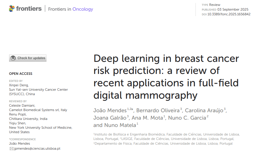
The recognition of her work by peers is evidenced by the invitation to review papers from international peer-reviewed journals in the medical imaging and artificial intelligence fields. In addition, she is member of the Editorial Board of “Frontiers in Oncology” as Review Editor for Radiation Oncology (Q2).
In addition to the research projects she is involved in the field of medical imaging, since 2022 she has been Invited Assistant Professor at the same Faculty, teaching subjects for Bologna Bachelor and Bologna Master Degrees in Biomedical Engineering and Biophysics.
Publicações
Publicações em revistas
(2023) Digital Breast Tomosynthesis: Towards Dose Reduction through Image Quality Improvement, Journal of Imaging 9(6), p. 119, MDPI, doi:10.3390/jimaging9060119
(2022) Automatic Classification of Simulated Breast Tomosynthesis Whole Images for the Presence of Microcalcification Clusters Using Deep CNNs, Journal of Imaging 8(9), p. 231, MDPI, doi:10.3390/jimaging8090231
(2020) Impact of total variation minimization in volume rendering visualization of breast tomosynthesis data, Computer Methods and Programs in Biomedicine 195, p. 105534, Elsevier, doi:10.1016/j.cmpb.2020.105534
(2020) Optimization of Breast Tomosynthesis Visualization through 3D Volume Rendering, Journal of Imaging 6(7), doi:10.3390/JIMAGING6070064
(2020) An Enhanced Visualization of DBT Imaging Using Blind Deconvolution and Total Variation Minimization Regularization, IEEE Transactions on Medical Imaging 39(12), p. 4094-4101, IEEE, doi:10.1109/TMI.2020.3013107
(2016) Dynamic relaxation in algebraic reconstruction technique (ART) for breast tomosynthesis imaging, Computer Methods and Programs in Biomedicine 132, p. 189-196, Elsevier, doi:10.1016/j.cmpb.2016.05.001
(2015) Total variation minimization filter for DBT imaging, Medical Physics 42(6), p. 2827-2836, doi:10.1118/1.4919680
Publicações em conferências
(2022) Detection of Microcalcifications in Digital Breast Tomosynthesis using Faster R-CNN and 3D Volume Rendering, PROCEEDINGS OF THE 15TH INTERNATIONAL JOINT CONFERENCE ON BIOMEDICAL ENGINEERING SYSTEMS AND TECHNOLOGIES (BIOIMAGING), VOL 2, p. 80-89, doi:10.5220/0010938800003123
(2020) Calculation of transfer functions for volume rendering of breast tomosynthesis imaging, 15th International Workshop on Breast Imaging (IWBI2020) 11513, p. 12, doi:10.1117/12.2559932
(2018) The underrated dimension: How 3D interactive mammography can improve breast visualization, Lecture Notes in Computational Vision and Biomechanics 27, p. 329-337, doi:10.1007/978-3-319-68195-5_36
(2017) Implementation of maj orization-minimization (MM) algorithm for 3D total variation minimization in DBT image reconstruction, 2016 IEEE Nuclear Science Symposium, Medical Imaging Conference and Room-Temperature Semiconductor Detector Workshop, NSS/MIC/RTSD 2016 2017-Janua, p. 1-5, doi:10.1109/NSSMIC.2016.8069493
(2016) An iterative algorithm for total variation minimization in DBT imaging, Computational Vision and Medical Image Processing V - Proceedings of 5th Eccomas Thematic Conference on Computational Vision and Medical Image Processing, VipIMAGE 2015(VipIMAGE 2015, Tenerife, Spain, October), p. 119-122, doi:10.1201/b19241-21
(2016) 3D total variation minimization filter for breast tomosynthesis imaging, Lecture Notes in Computer Science (including subseries Lecture Notes in Artificial Intelligence and Lecture Notes in Bioinformatics) 9699, p. 501-509, doi:10.1007/978-3-319-41546-8_63
(2015) Erratum to: Establishment of an open database of realistic simulated data for evaluation of partial volume correction techniques in brain PET/MR(EJNMMI Physics), EJNMMI Physics 2(1), p. 1, doi:10.1186/s40658-015-0129-9
Capítulos de livros
(2016) Digital breast tomosynthesis: A review of population-based screening/clinical trials, Digital Tomosynthesis: Benefits, Clinical Uses and Limitations, p. 25-74, Nova Science Publishers
Dissertações/Teses
(2010) Minimização do Ruído em Imagens de Mamografia por Emissão de Positrões através da Optimização do Tempo de Aquisição e do Tamanho de Voxel


